Guidelines classic scabies: in children < 15kg, pregnant or breastfeeding women
Updated on Jan 24
Responsibility
The information provided by this website comes from sources deemed reliable. However, the Société Française de Dermatologie recommends that the user ensure the validity of this information. Some may prove to be erroneous or be subject to typos or display errors.
The use of this data is under the sole responsibility of the user. The Société Française de Dermatologie cannot be blamed for a misinterpretation of the data provided by the site, or in the event of erroneous information. This decision tree and all the contents of this site have been developed in the context of updated data from science according to the HAS methodology, expert opinions and reviewers of the various documents and in the context of the French healthcare system.
The diagnosis of classic scabies is made during the patient's examination by the doctor.
The main symptom is the presence of chronic, almost constant itching (pruritus), which intensifies at night. The presence of itching in family members and in the environment can guide the diagnosis.
Scabies, known as "clean people's scabies," is often challenging to diagnose due to the rarity of lesions. It should be considered in the presence of persistent diffuse chronic itching.
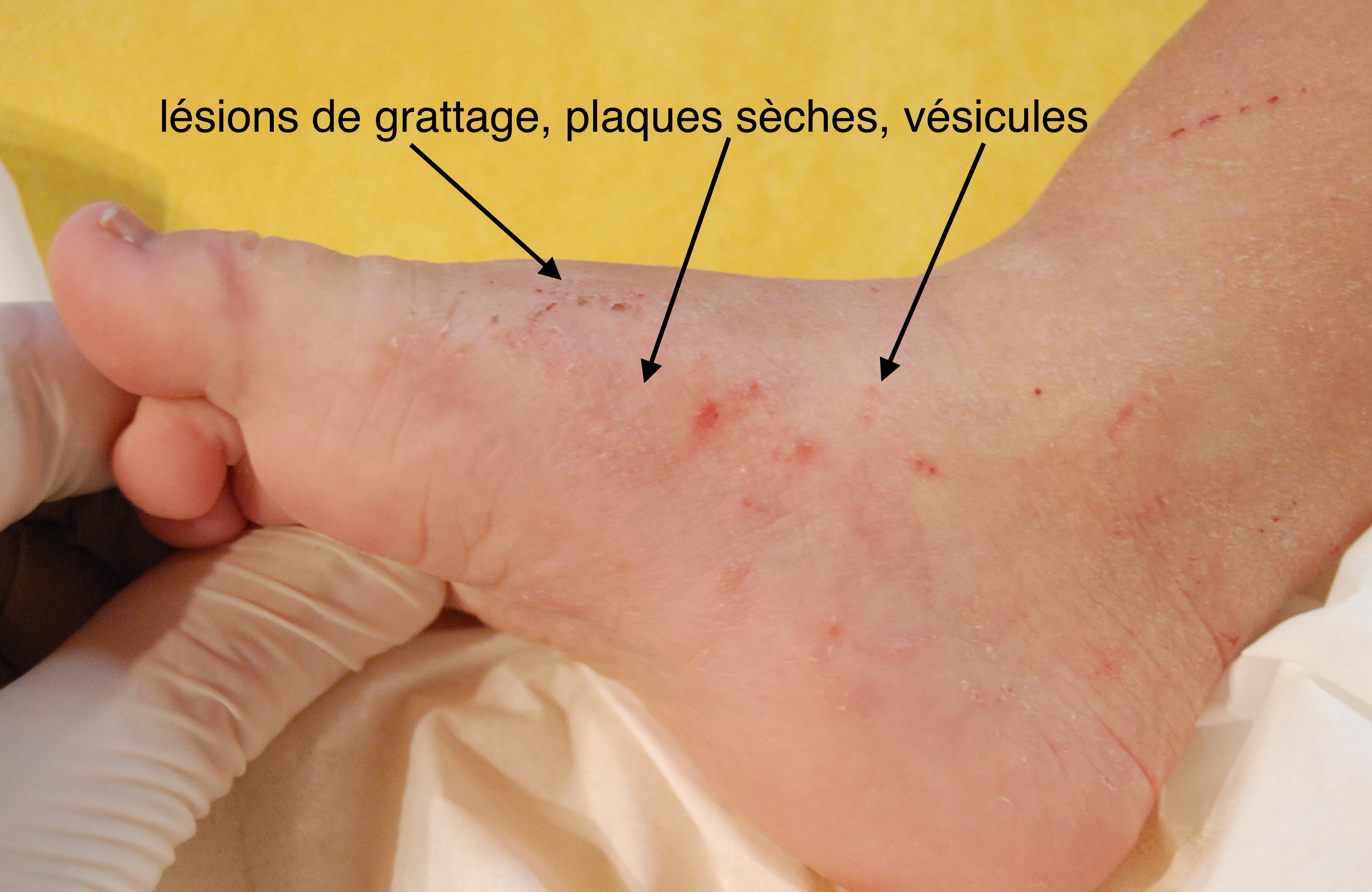
The most common skin lesions are not specific as they are related to scratching or the body's inflammatory immune reaction to the presence of the sarcoptes in the skin: they include linear streaks, scratches, small crusts, red patches, and dryness of the skin.
The specific lesions are not always present and should be carefully sought:
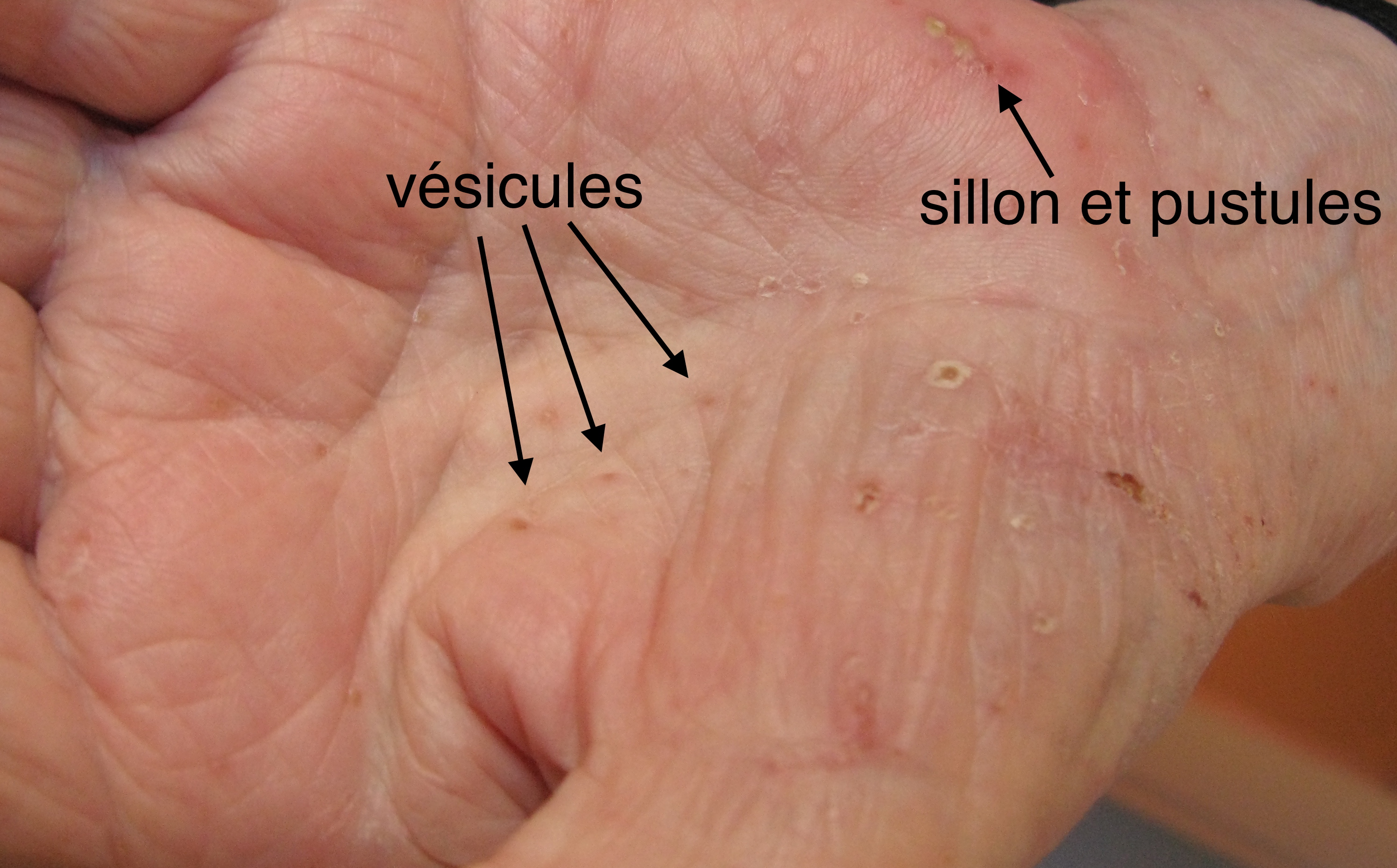
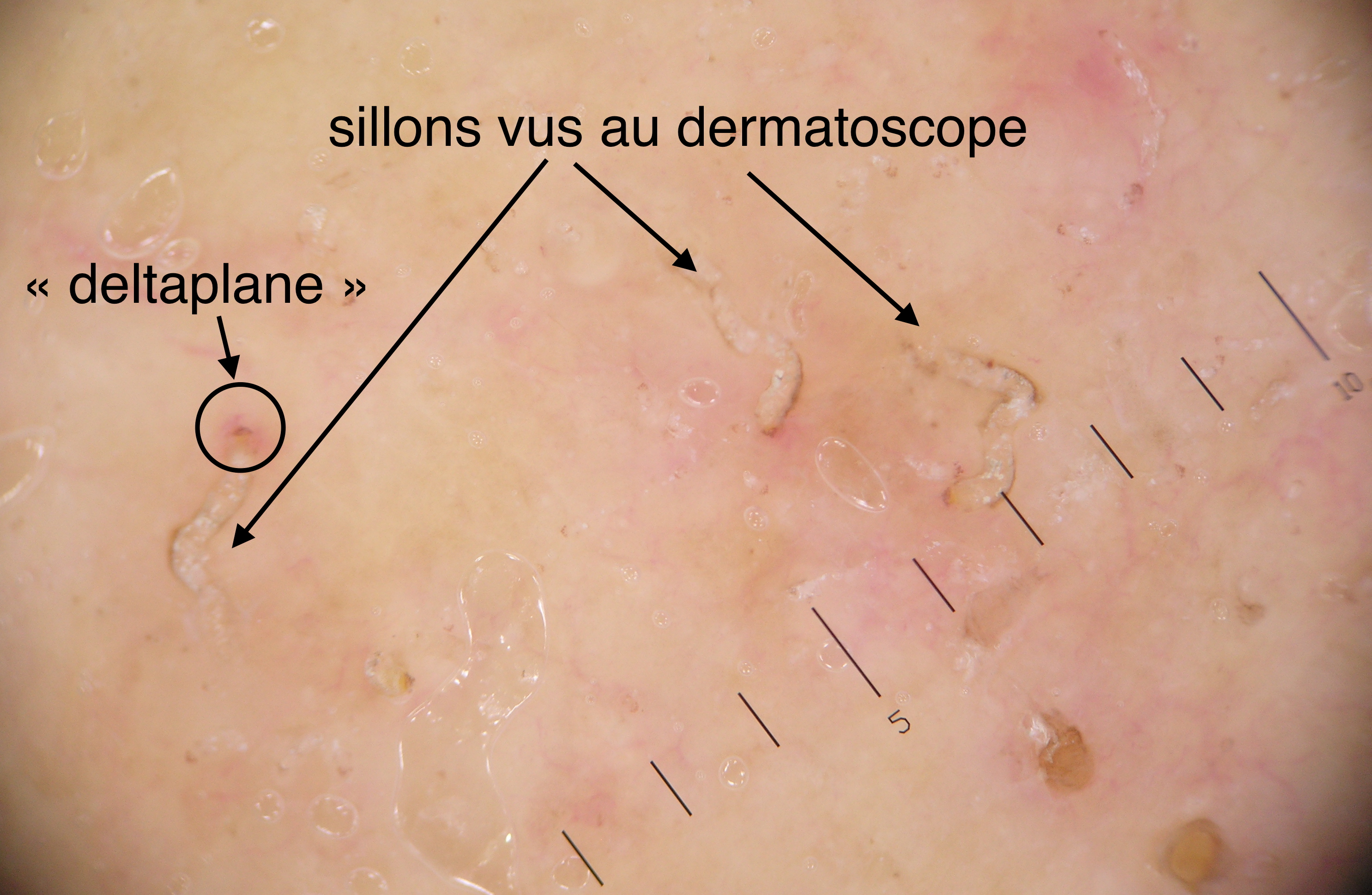
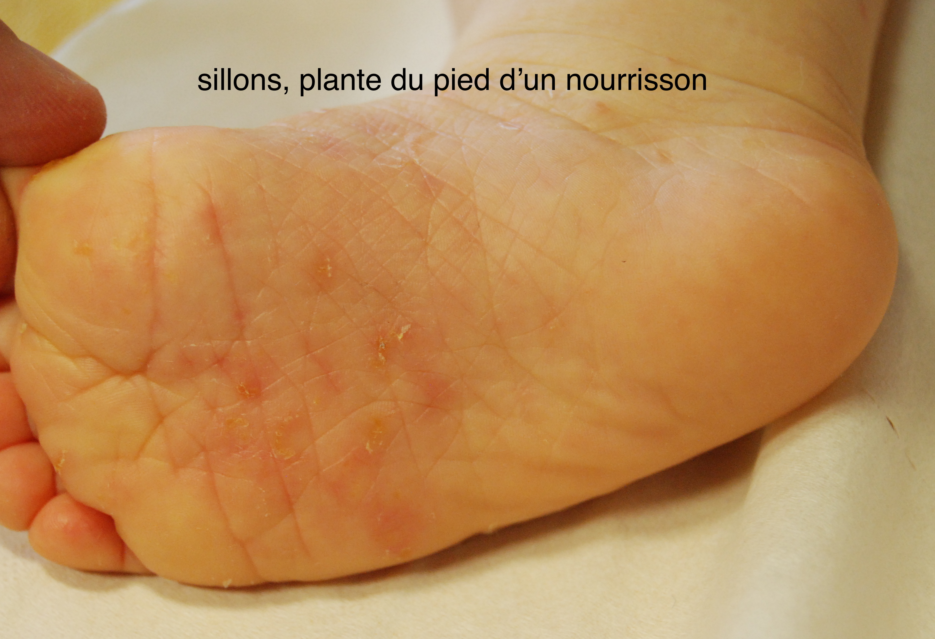
The burrows: Lesions a few millimeters in the form of sinuous lines, due to the parasite's path in the horny layer of the epidermis.
The pearl vesicles: Small elevations or tiny "blisters," the size of a pinhead, located at the end of the burrow (where the parasite resides).
Scabies Burrows
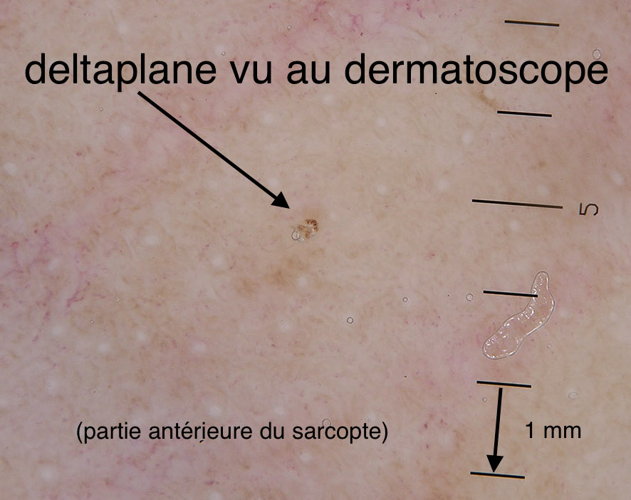
It is at the level of the vesicles and burrows that the parasite can most often be observed, using a dermatoscope placed on the skin. The sarcoptes are invisible to the naked eye, but under the dermatoscope, they appear as a very small black or dark brown triangle, resembling a hang glider or a Chinese hat, characteristic, and confirming the diagnosis (this hang glider corresponds to the anterior part of the parasite).
However, a negative search does not eliminate the diagnosis, especially when the number of parasites in the skin is low.
Scabies Burrows Hang glider, anterior part of the parasite
The other specific lesions are scabious nodules, red or purplish nodular lesions, raised, in domes, of several millimeters, or even centimeters, which are frequent especially in the scrotum in men. They are of immuno-allergic origin.
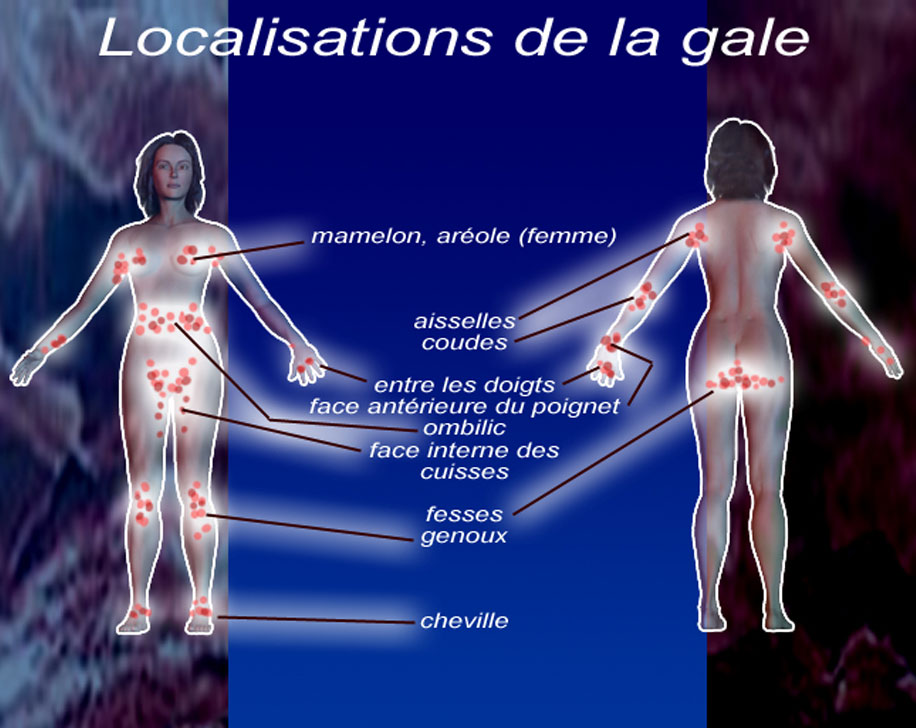
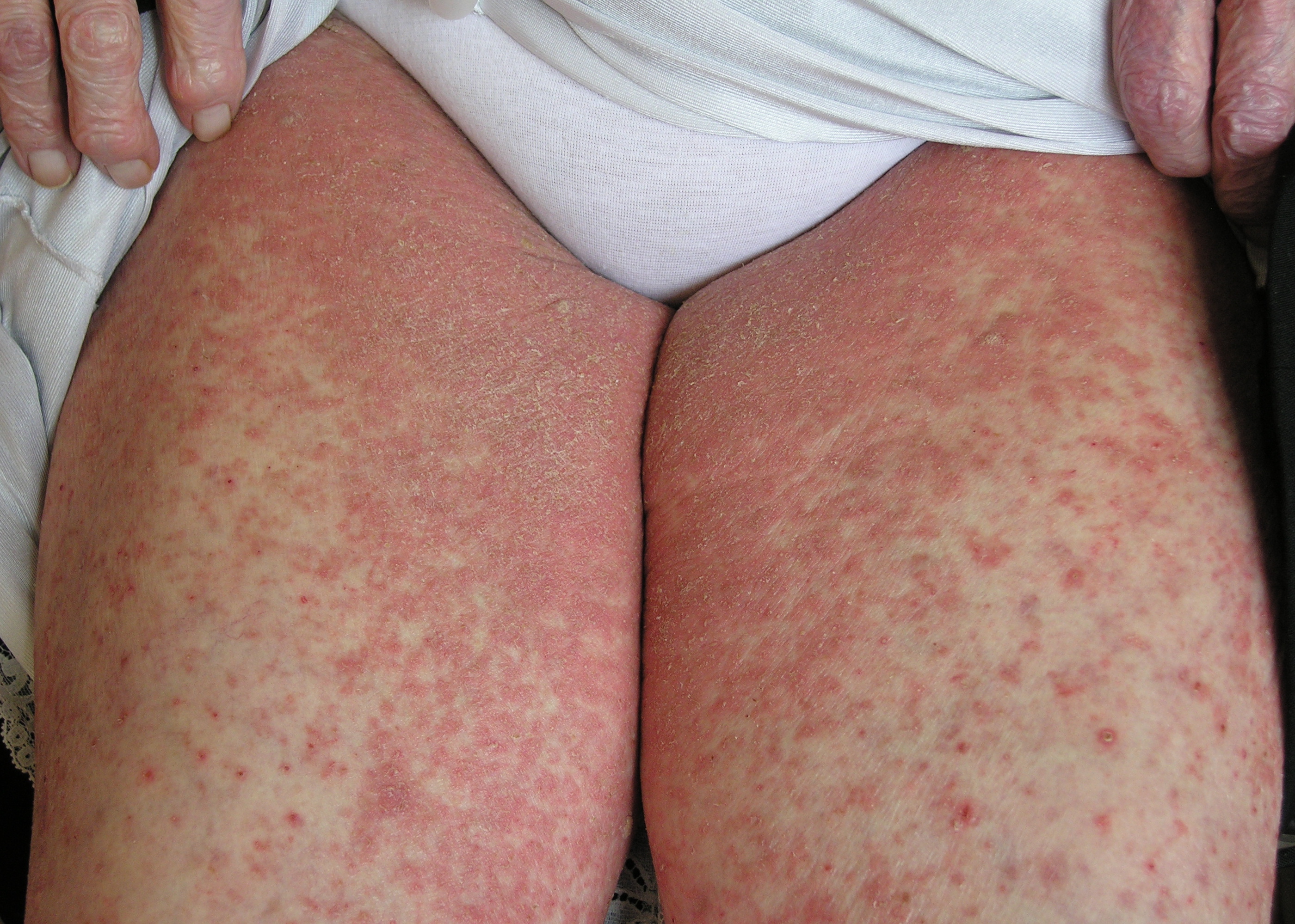
Location of lesions of Scabies
The topography of these lesions (specific and nonspecific) is suggestive of the diagnosis when they are located in the interdigital spaces (between the fingers), on the anterior surface of the wrists, at the elbows, in the armpits, on the buttocks, around the navel, on the inner thighs, especially in the genital area in men, and on the breasts in women.
The back and face are usually spared.
Scabies Location
Forms of Scabies
Profuse Scabies
Profuse or extended scabies is characterized by more numerous lesions and the extension of lesions throughout the body. Skin lesions can be found on the back. It is often associated with a late diagnosis, inappropriate treatments, or the presence of an unfavorable immune system, with decreased immunity.
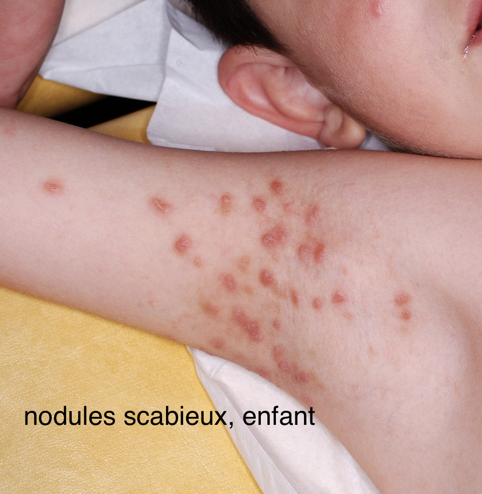
Infantile Scabies
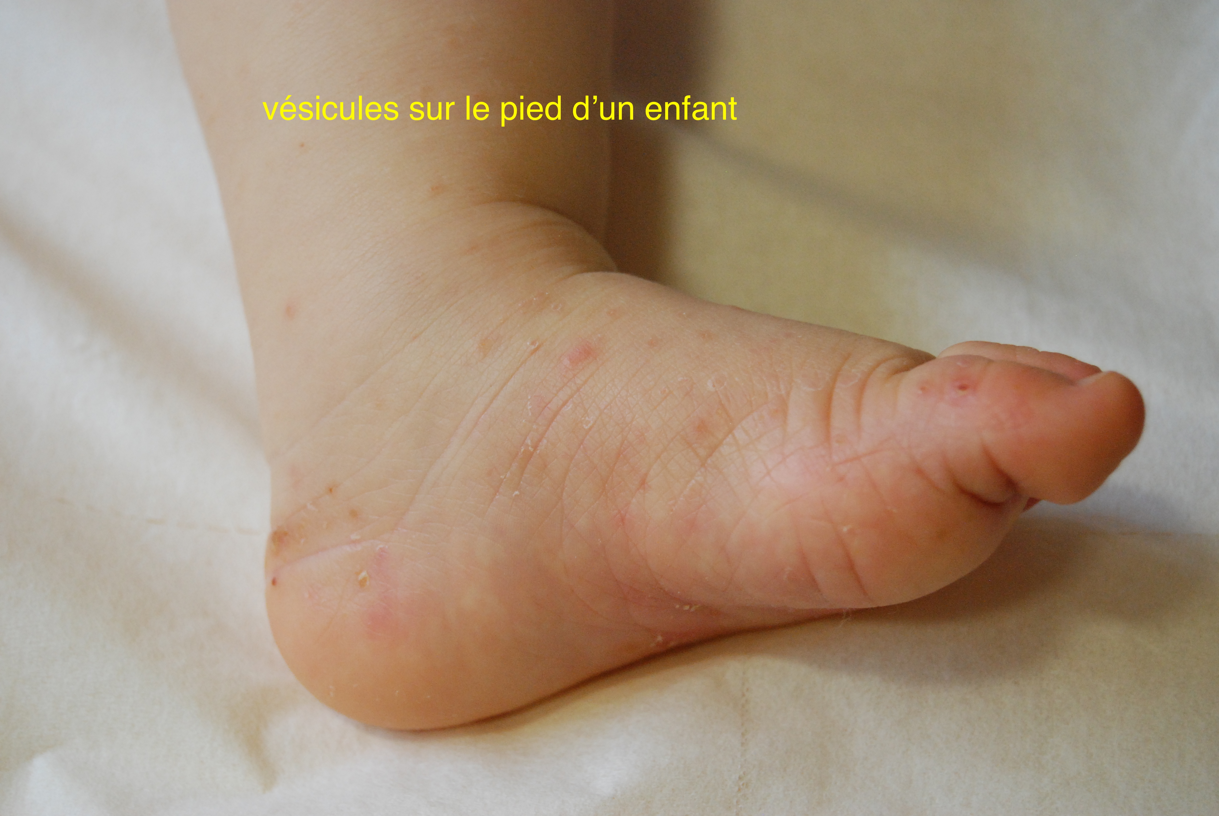
In very young children, scabies can have a deceptive appearance, including facial involvement, scabious nodules around the armpits, and pearl vesicles (small clear fluid blisters) or pustules (small turbid fluid blisters) on the palms of the hands and soles of the feet.
Scabies, scabious nodule in a child Scabies in a child Scabies, vesicles in a child Scabies, burrows in a child
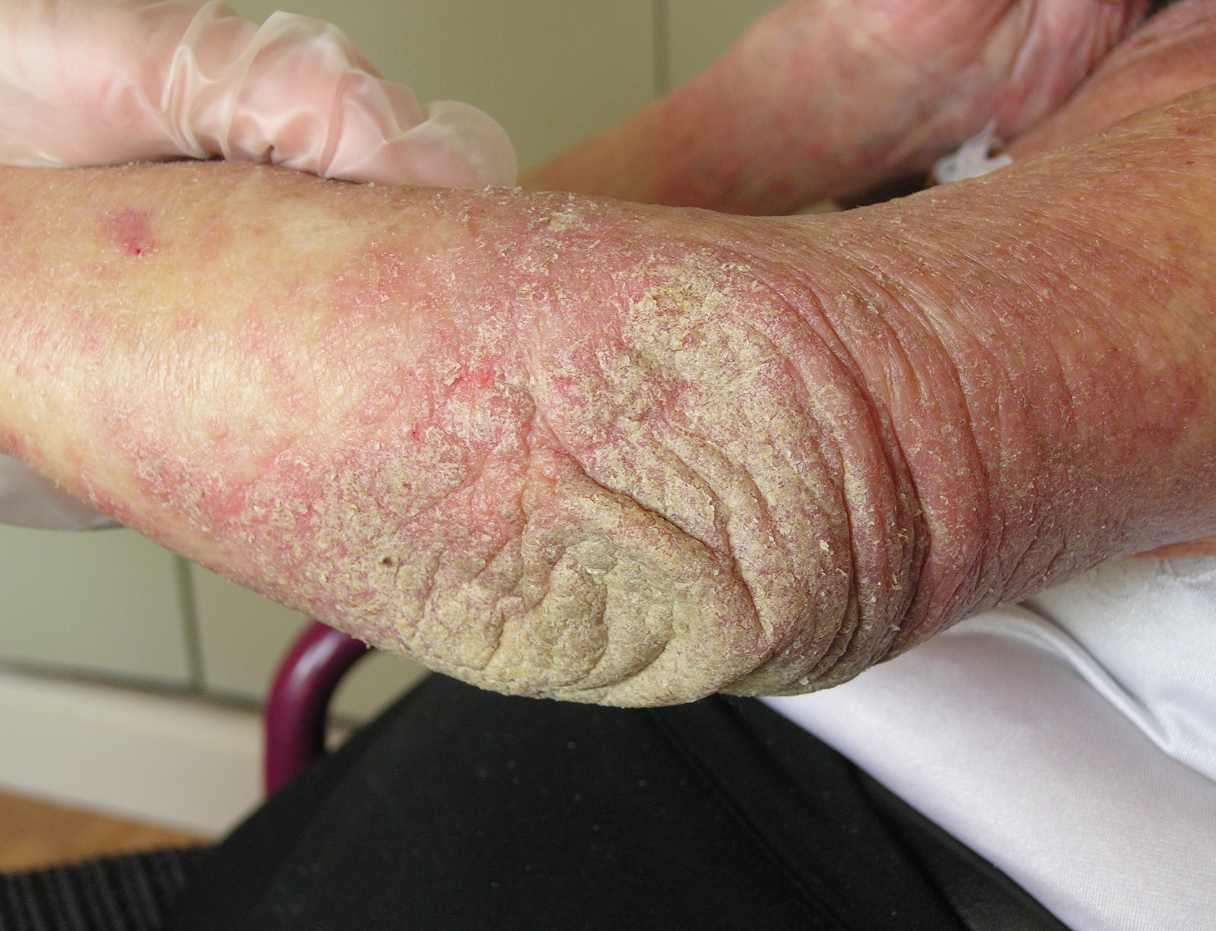
Hyperkeratotic Scabies (or Crusted Scabies)
It occurs in a specific context of immunosuppression (decreased natural defenses, secondary to a disease), or in elderly individuals living in a community setting.
The entire body is affected, including the face, scalp, and nails. There is erythroderma (diffuse redness of the entire body) with hyperkeratotic lesions, meaning that the horny layer of the skin is very thickened with scales and crusts, extensively. Importantly, itching may be absent or moderate.
Much less common than classic scabies, it is an extremely contagious form with thousands of parasites in the skin. Sometimes, hyperkeratotic scabies is limited to a skin segment.
Hyperkeratotic scabies Hyperkeratotic scabies
Complicated Scabies
Scabies lesions can be superinfected, especially by staphylococcus (bacteria that secondarily colonizes the skin affected by scabies parasites). This is referred to as impetiginization of the lesions. Eczema can also occur secondary to scabies, particularly in individuals with dry skin and/or intolerance to treatment.
Animal Scabies
There is also animal scabies related to other sarcoptes, including various types such as canine scabies, sheep scabies, etc. These different forms of scabies can be transmitted to humans, who represent a dead-end host (cannot transmit it to another human). Animal scabies also manifests with itching.
This notice will not be published on this site, but only sent to the publication management. Your email will only be used to reply to you if we deem it necessary. No response will be sent to any request for medical advice via this form.